
In the intricate landscape of neurology, pinpoint accuracy and comprehensive evaluations form the cornerstones of effective patient care. Central to this is the often underappreciated yet invaluable parameter in neuro exams: pupil diameter measurement.
A simple yet profound assessment, it serves as a window into the brain’s complex workings, providing key insights into the patient’s neurological health. The significance of accurately measuring pupil diameter in neurological evaluations cannot be overstated, with far-reaching implications for diagnosing and managing many conditions.
The Neuroscience Behind Pupil Diameter Changes
The regulation of pupil diameter hinges on the harmonious balance between the parasympathetic and sympathetic branches of the autonomic nervous system. In the parasympathetic pathway, signals from the Edinger-Westphal nucleus prompt the pupillary sphincter muscle to contract, causing the pupil to constrict. Conversely, the sympathetic system induces dilation through a series of neural connections.
Disruptions in these pathways can lead to tell-tale changes in pupil diameter, rendering this a key indicator of a patient’s neurological state. Whether it’s an excessive dilation hinting at sympathetic overdrive or a sluggish constriction suggesting parasympathetic dysfunction, the diameter of the pupils can be an essential signpost guiding a clinician’s assessment.
Importance of Pupil Diameter Measurement in Neuro Exams
In neuro exams, measuring pupil diameter is not merely a routine task; it is an insightful practice that can reveal much about a patient’s neurological function. Through this measurement, clinicians can glean information about the functionality of the autonomic nervous system and thereby speculate about potential neurological disorders.
For example, a fixed dilated pupil could indicate raised intracranial pressure, while unequal pupils could suggest a local mass effect or a systemic condition. The precise pupil diameter measurement is integral to neurological evaluations.
Common Methods of Pupil Diameter Measurement
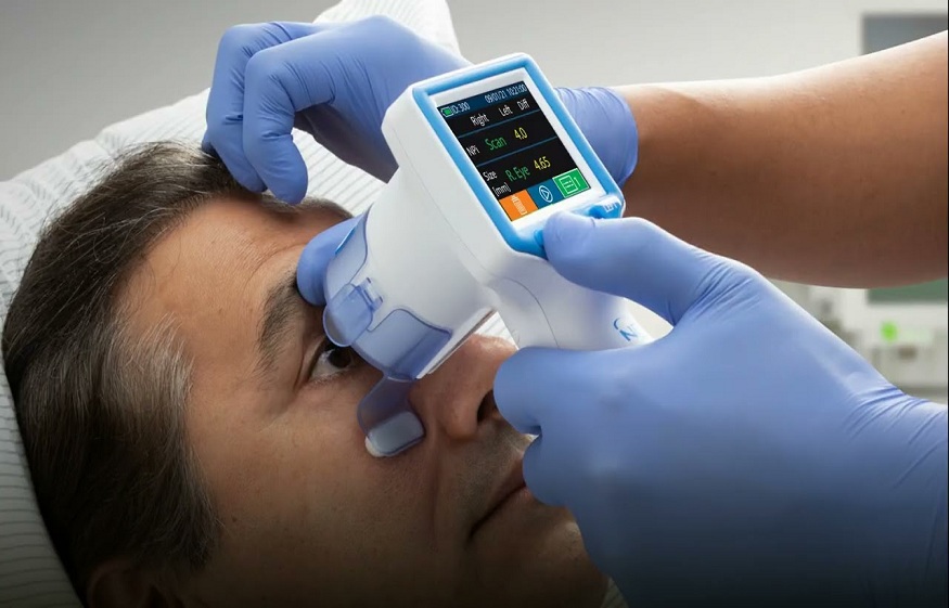
Two main methods dominate pupil diameter measurement. The traditional, manual method involves visual assessment using a penlight and a pupillary gauge. Although easily accessible and straightforward, this method suffers from potential inaccuracies and inter-examiner variability. In contrast, automated devices such as pupillometers offer high precision, eliminating the subjectivity inherent in manual measurements.
Cutting-edge neurological tools, like the NPi pupilometer, take this a step further by quantifying pupillary reactivity and size, enhancing the depth of pupil evaluation. However, these advanced methods come with challenges, including cost and the need for specific training.
Evaluation Criteria for the Pupil Diameter
Pupillary evaluation is typically based on three core parameters: size, shape, and reactivity. Each pupil’s size is expected to be between 2mm and 5mm under normal light conditions, with symmetry in size and shape between the two eyes.
Furthermore, both pupils should exhibit prompt constriction in response to light. Deviations from these norms, whether a size disparity between the two pupils or a sluggish light reaction, can signal potential neurological issues requiring further investigation.
Pupil Diameter Measurement and Brain Injuries
The relationship between pupil diameter measurement and brain injuries is well-established. The pupils often serve as the first sign of potential harm. For instance, an expanding brain lesion may manifest as anisocoria, a condition where the pupils are unequal.
Likewise, a dilated, unresponsive pupil may suggest impending brain herniation, a life-threatening condition. These examples underscore pupil diameter measurement’s critical role in diagnosing and managing brain injuries.
The Influence of Light on Pupil Diameter (100 words)
The pupil’s diameter inherently responds to ambient light conditions, constricting in bright light to limit light influx and dilating in low light to enhance visibility. Therefore, examining the pupils under different light intensities during a neuro exam is essential.
A lack of appropriate constriction or dilation could suggest autonomic dysfunction or a defect in the optic nerve pathway.
Equipment and Techniques for Pupil Diameter Measurement
Accurate pupil diameter measurement requires specialized equipment, from simple handheld light sources to advanced pupillometers. The Neurological Pupil Index (NPi) and the automated pupillometer have revolutionized this domain, providing precise diameter measurements and quantifying pupillary reactivity.
For accurate measurements, ensure a controlled light environment, maintain a consistent distance from the patient, and allow the pupils to adjust to the light source.
Conclusion:
In the intricate world of neuro exams, pupil diameter measurement plays a pivotal role. Its importance resonates with every neurological condition, from identifying brain injuries to diagnosing autonomic dysfunction. Through comprehensive pupil evaluations and the astute interpretation of findings, medical professionals can unlock a wealth of information hidden in the brain.